A happily pregnant mother arrived at the hospital to give birth to her baby boy. It was supposed to be one of those routine births, like many of the thousands that occur in each hospital every year. All the prenatal checks and tests during pregnancy suggested that everything was fine. Everything was going as expected, until the baby was born. At birth, he was blue, floppy, without a pulse, and not breathing. The midwife shouted for assistance and the medical team rushed over to help the baby breathe and to restart its heart. After battling for twenty minutes, the baby finally started to take its first independent breaths, before being sent to the neonatal intensive care unit. In spite of these efforts, brain damage had already begun.
This story is not unique. Approximately four in 1000 babies from developed countries are born with brain damage due to a lack of blood and oxygen immediately before birth. In developing countries, this number can be as high as one in five babies. This condition is known as hypoxic-ischaemic encephalopathy (HIE), and will often develop into cerebral palsy, causing difficulties in holding, walking, talking, and even swallowing.
Despite it being very common, HIE is still shrouded in mystery. In some cases, it arises as a consequence of a rupture in the placenta or in the uterus, a knot in the umbilical cord, or an infection. But for the majority of cases, the cause is unknown.
Newborn babies with HIE can be classified into three groups: mild, moderate and severe. This is assessed based on the severity of clinical symptoms such as floppiness of the body, level of heart rate and breathing, blueness of the skin, and excessive acid in the blood. For the moderate to severe HIE babies, the only treatment consists of cooling the baby’s body down to around 33 degrees Celsius for three consecutive days. For this treatment to work, it must be carried out within six hours of brain injury. However, this decision becomes very difficult to make when doctors are unsure when or how the brain damage occurred. In addition, this method will only work for one in nine babies.
To date, the most common way to check for brain damage is by doing an MRI scan. Again, this method has revealed to be unreliable due to the fact that brain injuries develop with time, and are more clearly visible between one and two weeks after birth. The most effective way of determining the severity of brain injury is two years after injury, using tests which focus on the child’s ability to walk, pick up small objects, speak, and understand basic questions. These methods, while popular, still fail at detecting brain injury before the six hour window has passed.
A more valid method of assessing the severity of brain damage immediately after birth is to do a brain biopsy, which involves extracting and examining a small bit of brain tissue. However, this raises an ethical problem, as in certain cases it can cause further brain damage. Since every part of the brain has an important function, the removal of any part, however small, will cause some damage to the brain. Furthermore, current understanding suggests that when the brain is damaged, it will never return to normal state. Scientists therefore have to identify a less invasive method of assessing brain damage.
Recently, scientists have tried to assess the severity of brain damage by looking at the contents of brain cells. When the brain lacks oxygen or blood, cells in the brain such as nerve and glial cells will start to die and break up, releasing their cell content. This causes the molecules which form the structure of the nerve cell to spill out into the surrounding cerebrospinal fluid (CSF), a natural fluid that bathes the brain. When this happens, it can be detected. Molecules in the CSF can therefore serve as ‘biological markers’ (or biomarkers for short), and can act as clues as to the severity of the brain injury.
 Blood samples from newborns could be used to diagnose and treat early brain injury
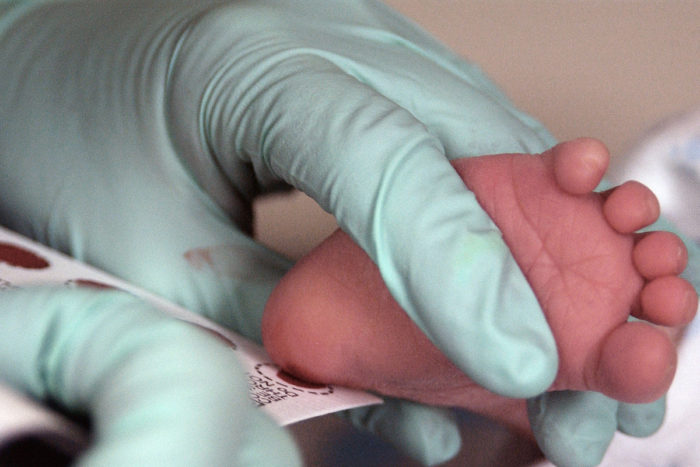
However, trying to get CSF from a sick baby is very difficult in practice, so scientists have looked at easier ways. Molecules in the CSF can also leak into the blood. The latest technologies allow scientists to work with amounts as small as a single drop of blood, from which they have successfully studied different types of molecules such as nucleic acids, proteins and lipids. This minimally invasive approach is now used routinely to screen all newborn babies for conditions such as sickle cell disease, cystic fibrosis, and metabolic diseases.
Recently, ethical approval has been obtained to set up the Brain Injury Biomarkers in Newborns Study. The initial steps of this study have involved five large specialized hospitals in England. Blood samples from babies with moderate-severe brain injury were collected at 3 time points; at the start, during, and after the cooling treatment. The molecules in these samples will be compared to those of blood belonging to babies with mild brain injury that did not require cooling treatment, or umbilical cord blood of healthy babies without brain injury. The data obtained from this study will help to identify which molecule or panel of molecules are related to brain injury.
A potentially significant molecule is the neurofilament light protein. This molecule is found only in nerve cells, used as structural support in the cell’s communication system. Recently, it has been shown that in babies with brain damage, this neurofilament light protein from damaged nerve cells in the brain is present in the blood. Furthermore, it has been found that the more brain damage the baby had according to a brain MRI scan, the more neurofilament light protein was present in their blood.
Research into the significance of other molecules including phospholipids, the main components of cell membranes, is still in its infancy and is currently ongoing.
With more research, scientists may soon be able to identify specific brain cell molecules in the blood that could be used to assess whether a newborn brain is damaged, as well as to determine the extent of the damage. Newborn life requires extremely delicate handling, and time in such situations is extremely precious. The success of biomarker studies will allow doctors to quickly decide which treatment to provide and track its progress. In the future, we may be able to save more newborns from lifelong afflictions that stem from the hurdles they face in their first hours of life.